
A Guide to Cellular Energy, Aging, and Recovery
An accessible introduction to what mitochondria are, how they support cellular function, why they change over time, and how they affect tissues throughout the body.
A foundation for understanding mitochondria
This guide begins with the basic structure and function of mitochondria, then moves into their origins, abundance, role in aging, and importance throughout the body.
Read the chapters in order or use the contents to return directly to a subject you want to study in greater detail.
Technical terms are introduced gradually, with definitions, illustrations, key points, and links to related articles for further reading.
What Are Mitochondria?
Mitochondria are specialized structures inside nearly all human cells. Their best-known role is converting energy from nutrients into a form that cells can use.
Every cell must perform work. Muscle cells contract, nerve cells transmit signals, kidney cells move substances, and other cells build, repair, transport, and communicate.
Much of this work depends on adenosine triphosphate, usually called ATP. ATP is a molecule that transfers usable energy within cells.
Mitochondria help produce ATP by processing energy from carbohydrates, fats, and other nutrients. In many cell types, this process uses oxygen and takes place across the mitochondrion’s highly folded inner membrane.
Mitochondria do more than produce ATP. They also participate in metabolic regulation, calcium signaling, controlled stress responses, cellular maintenance, and programmed cell death.
Anatomy at a glance
The main parts of a mitochondrion
A mitochondrion contains several distinct compartments. Each creates the physical conditions needed for particular chemical reactions and cellular processes.
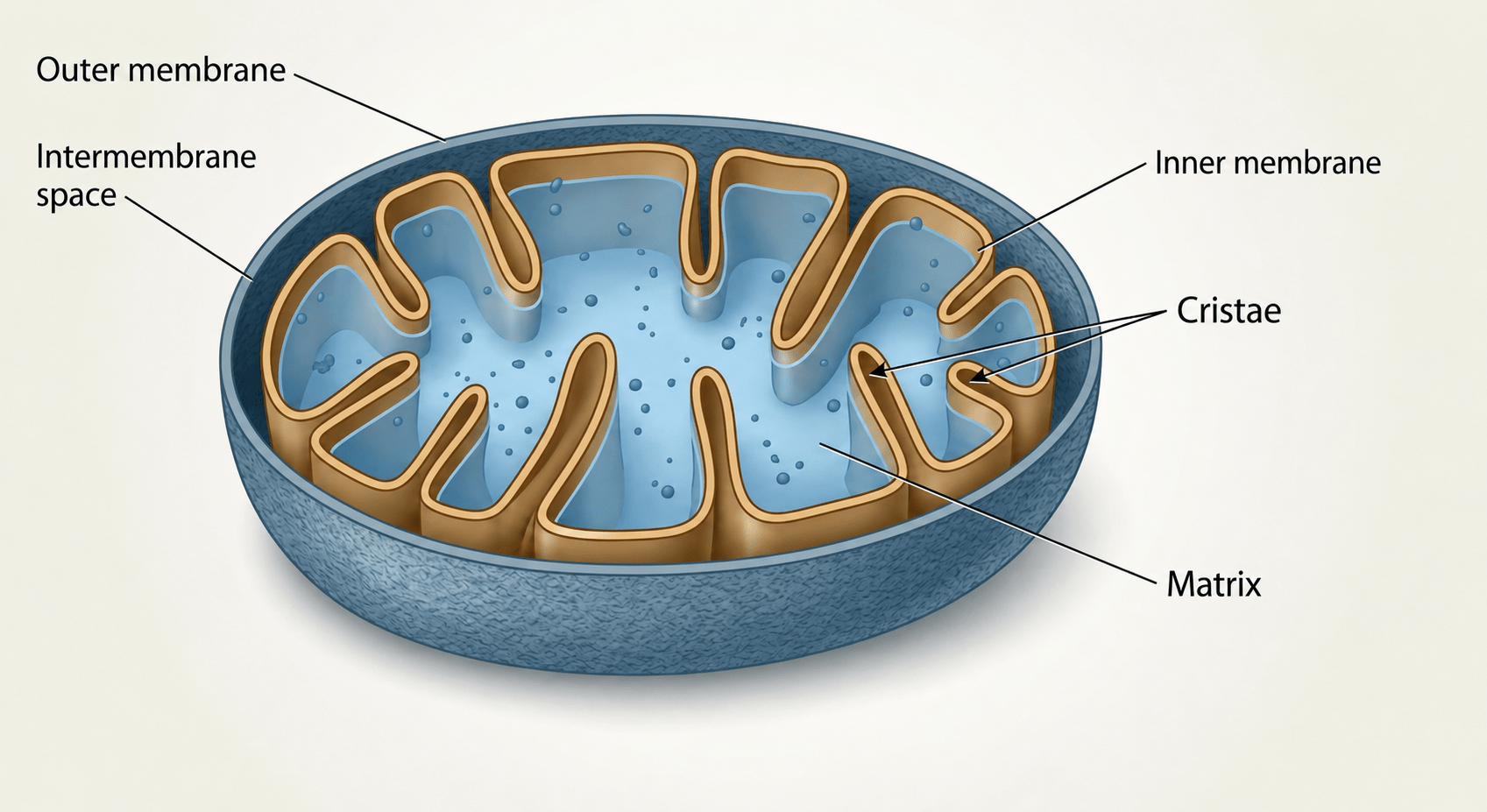
- Outer membrane
- The outer boundary of the mitochondrion. It separates the organelle from the surrounding interior of the cell.
- Intermembrane space
- The narrow compartment between the outer and inner membranes. It plays an important role in establishing the proton gradient used during ATP production.
- Inner membrane and cristae
- The inner membrane contains much of the machinery involved in oxidative phosphorylation. Its folds, called cristae, increase the available membrane area.
- Matrix
- The innermost compartment. It contains enzymes, mitochondrial genetic material, and components needed for several metabolic reactions.
A distinctive organelle
Mitochondria contain their own DNA
Most human DNA is stored in the cell nucleus. Mitochondria also contain a small amount of their own genetic material, known as mitochondrial DNA.
Mitochondrial DNA provides instructions for a limited number of components needed by the organelle. Most mitochondrial proteins, however, are encoded by nuclear DNA and transported into the mitochondria after they are produced.
This unusual arrangement is connected to the evolutionary origin of mitochondria, which is examined in the next chapter.
Where Did Mitochondria Come From?
Mitochondria appear to have begun as free-living bacteria that entered into a lasting biological partnership with an ancestral host cell.
The most widely accepted explanation for the origin of mitochondria is known as the endosymbiotic theory.
According to this theory, an ancestral cell engulfed an aerobic bacterium, a microorganism capable of using oxygen to extract energy from nutrients. Instead of being digested, the bacterium remained alive inside the host.
The relationship benefited both organisms. The bacterium gained a protected environment and a steady supply of nutrients, while the host gained access to highly efficient aerobic energy production.
Over a very long period of evolution, the two became increasingly interdependent. The bacterium gradually lost much of its ability to live independently and became the organelle now known as the mitochondrion.
The proposed sequence
From bacterium to organelle
The endosymbiotic theory describes a gradual evolutionary transition, not a single event that produced a modern mitochondrion immediately.
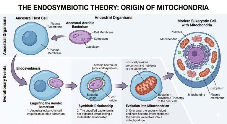
Engulfment
An ancestral host cell enclosed an aerobic bacterium within its cell membrane.
Stable partnership
The bacterium survived inside the host, receiving protection and nutrients while helping the host use oxygen more efficiently.
Evolutionary integration
Over time, the bacterium transferred or lost many genes and became dependent on the host cell, eventually functioning as an organelle.
Evidence
Why scientists accept this explanation
- Mitochondria contain their own DNA. Mitochondrial DNA is separate from the DNA stored in the cell nucleus.
- Their DNA is circular. This resembles the organization of bacterial chromosomes more than the linear chromosomes found in the human nucleus.
- They are surrounded by two membranes. This is consistent with an ancestral bacterium being engulfed by another cell.
- They divide by a fission-like process. Existing mitochondria grow and divide rather than being assembled from nothing by the cell.
- Their protein-making machinery has bacterial features. Mitochondrial ribosomes differ from the ribosomes found in the main body of the human cell.
How Many Mitochondria Do We Have?
There is no single mitochondrial count for the human body. The number, size, shape, and density of mitochondria differ greatly among cell types and can change as cellular energy demands change.
Some cells contain no mitochondria, while others contain hundreds, thousands, or far more. These differences reflect the specialized work performed by each type of cell.
Cells that perform continuous or energy-intensive work generally devote more space and resources to mitochondrial energy metabolism. Heart muscle cells, for example, must contract without interruption, while liver cells carry out a wide range of metabolic and detoxification processes.
Neurons also require substantial energy to maintain electrical gradients, transmit signals, and support communication across long cellular extensions. Within a neuron, mitochondria are often concentrated in areas with especially high energy demand.
Mature red blood cells are an important exception. They remove their mitochondria during development, leaving more space for hemoglobin and preventing the cells from consuming the oxygen they transport.
Comparative reference
Estimated abundance in selected human cells
These broad estimates illustrate how mitochondrial abundance can vary among different cell types and levels of cellular energy demand.
| Tipo de célula | Número aproximado de mitocondrias por célula | Notas |
|---|---|---|
| Glóbulos rojos (eritrocitos) | 0 | Mature erythrocytes lack mitochondria to maximize hemoglobin content and oxygen transport capacity. |
| Glóbulos blancos (leucocitos) | 3–100 | Typically low at rest, as in lymphocytes, and higher in more metabolically active or activated immune cells. |
| Células epiteliales | 100–500 | Moderate abundance that varies by tissue and transport workload. Renal tubular epithelial cells, for example, generally have higher mitochondrial content. |
| Neuronas | 100–2,000 | Variable and often concentrated in regions with high ATP demand, including synapses, nodes, and long axons. |
| Células del hígado (hepatocitos) | 800–2,000 | High abundance reflecting intensive metabolic activity, biosynthesis, and detoxification pathways. |
| Fibras musculares esqueléticas | 200–5,000+ | Highly variable and influenced by muscle-fiber type, activity level, and training status. Endurance training can increase mitochondrial density. Learn more about how exercise changes mitochondrial capacity. |
| Células del músculo cardíaco (cardiomiocitos) | 3,000–8,000 | Very high abundance to support continuous contraction, ion transport, and cellular maintenance. |
| Oocitos (óvulos) | 100,000–600,000+ | Extremely high abundance to help support oocyte maturation, fertilization, and early embryonic development before robust mitochondrial biogenesis begins. |
| Otras células somáticas (rango habitual) | 100–1,000 | A common estimated range for many non-specialized cell types, including fibroblasts. |
Values are approximate and vary by tissue type, physiological state, species, and measurement method.
Interpreting the numbers
More mitochondria does not automatically mean better function
Mitochondrial abundance is only one part of cellular energy biology. A cell can contain many mitochondria that function poorly, or a smaller population that operates efficiently and responds well to changing demand.
Researchers therefore examine several related features, including mitochondrial volume, structure, respiratory capacity, membrane organization, and the balance between mitochondrial formation, fusion, division, repair, and removal.
For a practical look at how these systems can change over time, see how long it takes to improve mitochondrial capacity.
Tipo de célula
Different tissues have different energy requirements and therefore maintain different mitochondrial populations.
Energy demand
Repeated or sustained cellular work can influence mitochondrial content and metabolic capacity.
Fusion and fission
Mitochondria join, divide, and remodel their networks in response to cellular conditions. These changes work alongside mitochondrial biogenesis and mitophagy, the processes that help cells renew capacity and remove damaged mitochondria. Read more about mitochondrial biogenesis and mitophagy.
Age and health
Mitochondrial abundance, structure, and function can change with development, aging, disease, and environmental stress.
What Do Mitochondria Do?
Mitochondria are best known for producing ATP, but their role extends well beyond energy production. They also help cells regulate metabolism, respond to stress, manage calcium, communicate with the immune system, and determine when damaged cells should be removed.
Mitochondria are not passive structures that simply produce energy at a constant rate. They continually respond to changes in nutrient availability, oxygen, physical activity, cellular stress, and tissue demand.
A contracting muscle cell, an active neuron, and a liver cell processing nutrients place different demands on their mitochondria. The mitochondrial network adjusts its activity, structure, and fuel use accordingly.
This adaptability is one reason mitochondrial health cannot be reduced to a single measurement. ATP production matters, but so do mitochondrial quality control, redox balance, membrane organization, signaling, and the ability to respond to changing conditions.
The sections below introduce the principal functions of mitochondria. The process by which mitochondria produce ATP will be examined in greater detail in the next chapter.
Core functions
Six major roles of mitochondria
These functions are interconnected. A change in energy production can affect signaling, stress responses, metabolism, repair, and the ability of the cell to maintain itself.
ATP production
Mitochondria convert energy from nutrients into ATP, the molecule cells use to power muscle contraction, ion transport, biosynthesis, movement, communication, and repair.
Fuel metabolism
Mitochondria help process carbohydrates, fatty acids, and certain amino acids. They adjust fuel use according to nutrient availability, activity, hormonal signals, and tissue demand.
Calcium regulation
Mitochondria take up and release calcium, helping cells coordinate processes such as muscle contraction, nerve signaling, secretion, metabolism, and stress responses.
Redox signaling
Mitochondria produce reactive oxygen species as part of normal metabolism. At controlled levels, these molecules participate in signaling and adaptation. In excess, they can contribute to oxidative stress.
Immune and stress signaling
Mitochondria help cells detect and communicate damage, infection, and metabolic stress. Their membranes, metabolites, reactive molecules, and DNA can all participate in immune signaling.
Cell survival and programmed cell death
When damage becomes too severe, mitochondria can help initiate apoptosis, an organized process that removes cells that are no longer safe or useful to the tissue.
A coordinated network
Mitochondria change with cellular demand
Mitochondria continually change their shape, position, and organization. They can join through fusion, divide through fission, move to areas of high energy demand, and remove damaged components through quality-control pathways.
These processes allow a mitochondrial network to redistribute materials, isolate damaged regions, adjust energy capacity, and respond to changes in the cellular environment.
Mitochondrial renewal depends on balance. Cells need mechanisms that build and maintain capacity, but they also need systems that identify and recycle mitochondria that are no longer functioning well. Our article on mitochondrial biogenesis and mitophagy explains how these processes work together.
Fusion
Mitochondria join and share membranes, proteins, metabolites, and genetic material within the network.
Fission
A mitochondrion divides, helping the network redistribute, expand, or isolate a damaged region.
Biogenesis
Cells produce new mitochondrial components and expand or renew mitochondrial capacity.
Mitofagia
Damaged or unnecessary mitochondrial material is selectively identified and recycled.
Related reading
Continue exploring mitochondrial function
Mitochondria and Aging
Aging affects the systems that produce, maintain, repair, and recycle mitochondria. These changes can gradually reduce cellular energy capacity and make tissues less resilient when demand increases.
Aging does not cause every mitochondrion to stop working at once. Instead, many small changes accumulate across the mitochondrial network over time.
Mitochondria may become less efficient, molecular damage may accumulate, and the systems responsible for removing damaged components and rebuilding capacity may become less responsive.
The result is often a reduction in mitochondrial reserve, the additional energy capacity cells can call upon when workload increases.
Biological changes
What changes in mitochondria as we age?
Aging can affect mitochondrial energy production, structural integrity, quality control, and responsiveness. These changes overlap and may reinforce one another.
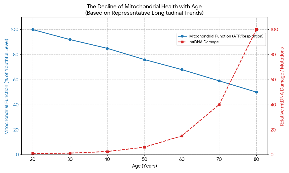
Reduced energy-producing capacity
Mitochondrial respiration and ATP production may become less efficient, particularly in energy-demanding tissues such as muscle, heart, and brain.
Accumulation of molecular damage
Changes can accumulate in mitochondrial DNA, proteins, lipids, membranes, and respiratory machinery after years of metabolic and environmental stress.
Weaker quality control
Fusion, fission, repair, and mitophagy may become less coordinated, allowing damaged or inefficient mitochondrial material to remain within the network.
Reduced renewal and adaptation
Signaling involved in mitochondrial biogenesis may become less responsive, limiting the ability to rebuild capacity when cellular demand rises.
Functional consequences
Why these changes matter
Reduced mitochondrial reserve may become most noticeable when the body is asked to perform, recover, or adapt under increased demand.
Lower stamina and muscle resilience
Familiar activity may require more effort when muscle has less oxidative capacity and less reserve available for sustained work.
Slower recovery and reduced stress tolerance
Tissue repair, ion balance, protein turnover, and adaptation all require energy. Poor sleep, illness, and inactivity may therefore have a larger effect.
Changes in cognitive and metabolic endurance
Neurons and metabolically active tissues depend on reliable energy supply, making sustained mental effort and fuel switching more difficult when reserve is reduced.
Adaptive capacity
Mitochondria remain responsive throughout life
Age-related mitochondrial decline is not identical in every person. Genetics, disease, physical activity, muscle mass, cardiovascular health, sleep, nutrition, medication, and environmental exposure all influence the trajectory.
Mitochondria also remain responsive to biological demand. Exercise is one of the best-studied signals for maintaining or improving mitochondrial capacity in skeletal muscle because it activates pathways involved in remodeling, oxidative metabolism, and biogenesis.
This does not mean every age-related change can be reversed. It means that biological aging and inactivity are not the same process, and that maintaining healthy demand can help preserve function.
What Happens When Mitochondria Don’t Function Properly?
When mitochondria cannot meet cellular demand efficiently, cells may have less usable energy, less metabolic flexibility, and a reduced ability to adapt, repair, and recover.
Mitochondrial function exists on a spectrum. A mitochondrion may continue producing energy while operating less efficiently, responding poorly to increased demand, or producing more stress signals than the cell can manage.
Problems become more important when impaired mitochondria accumulate or when repair and recycling cannot keep pace with damage.
High-demand tissues such as the brain, heart, skeletal muscle, liver, and kidneys may be especially sensitive because they depend on a steady energy supply and tightly regulated mitochondrial signaling.
Cellular consequences
Four ways reduced mitochondrial function can affect a cell
These effects are connected. Reduced energy can limit maintenance, while accumulated damage and stress can further interfere with mitochondrial performance.
Less usable energy
Cells may have less ATP available for contraction, transport, electrical signaling, biosynthesis, maintenance, and repair.
Reduced fuel flexibility
Cells may become less effective at processing fats and carbohydrates or switching between fuels as availability and demand change.
More cellular stress
Disrupted electron flow, calcium imbalance, membrane damage, and excessive reactive oxygen species can activate stress pathways.
Slower repair and recovery
When energy is limited, cells may prioritize immediate survival over protein renewal, tissue remodeling, adaptation, and long-term maintenance.
A reinforcing pattern
Dysfunction can create additional strain
Reduced mitochondrial function is rarely limited to ATP production. When less energy is available, repair and quality control may weaken. Damaged components can then accumulate and place further strain on the mitochondrial network.
Mitochondrial stress can also activate immune signaling. Persistent inflammation may interfere with respiration, membrane function, and mitochondrial recycling, creating a two-way relationship.
Interpreting symptoms
Mitochondrial dysfunction does not have one unique feeling
Reduced cellular capacity may be experienced as lower stamina, faster fatigue, slower recovery, reduced exercise tolerance, or difficulty sustaining physical or mental effort.
These experiences are not specific to mitochondria. They can also result from sleep disorders, anemia, thyroid conditions, infection, cardiovascular or respiratory disease, medication effects, insufficient nutrition, psychological stress, and many other causes.
Mitochondrial biology can help explain how cellular capacity affects function, but it cannot identify the cause of an individual symptom. A broader discussion is available in What Does “Mitochondrial Dysfunction” Actually Feel Like?
Mitochondria Throughout the Body
Every tissue depends on mitochondria, but not in exactly the same way. The number, organization, fuel use, and activity of mitochondria reflect the specialized demands of each cell type.
A heart cell must contract continuously. A neuron must maintain electrical gradients and communicate across long distances. A liver cell must process nutrients, manage fuel storage, and respond to changing metabolic conditions.
These different workloads shape how mitochondrial function is organized throughout the body. Tissues with high or rapidly changing energy demands often depend on substantial mitochondrial capacity and tightly regulated quality control.
The interactive map below introduces several major tissues and systems in which mitochondrial biology is actively studied.
Systems overview
How mitochondrial health connects across the body
Select a body area or system label to examine how mitochondrial function relates to its specialized biological demands.
Pulsing points and labels are interactive

Selected tissue or system
Neurology and Brain Health
The brain has high energy demands, and mitochondrial function is studied in relation to memory, focus, mood, cognitive aging, and cellular resilience.
Why mitochondria matter here
Brain cells depend heavily on mitochondrial activity to help manage energy demand, oxidative stress, cellular signaling, and synaptic function.
Integrated physiology
No tissue functions in isolation
Although each tissue has specialized mitochondrial demands, the systems shown above are closely connected. The heart and blood vessels deliver oxygen and nutrients. The liver processes and stores fuels. Hormones influence metabolic demand, while immune signaling changes how tissues allocate energy during stress or injury.
Skeletal muscle also acts as a major metabolic organ. Its activity influences glucose use, circulation, signaling, and whole-body energy balance. The brain coordinates many of these responses while depending on a continuous energy supply of its own.
How Can Mitochondrial Function Be Supported?
Mitochondria respond to the demands and conditions they experience. Movement, sleep, nutrition, recovery, and overall health influence how cells build, use, maintain, and renew mitochondrial capacity.
Supporting mitochondrial function is not based on one isolated intervention. Mitochondria adapt to repeated biological signals over time.
Some signals increase energy demand and encourage adaptation. Others provide the fuel, materials, and recovery conditions required for maintenance and renewal.
The central goal is to create a consistent environment in which cells can respond to demand without being overwhelmed by it.
Major influences
Four factors that shape mitochondrial capacity
These factors work together. Exercise creates demand, while sleep, nutrition, recovery, and overall health help determine whether that demand becomes useful adaptation.
Regular movement and exercise
Aerobic and resistance exercise increase cellular energy demand. Repeated training can stimulate mitochondrial remodeling, biogenesis, fuel use, and greater respiratory capacity in skeletal muscle.
Read Exercise and MitochondriaSleep and circadian regularity
Sleep supports metabolic regulation, hormonal balance, cellular maintenance, and recovery. Irregular or insufficient sleep can disrupt the conditions in which adaptation occurs.
Nutrition and energy availability
Cells need adequate energy, protein, essential fats, vitamins, and minerals to maintain membranes, enzymes, antioxidant systems, and other components involved in mitochondrial function.
Read Food, Fasting, and MitochondriaRecovery and overall health context
Stress, inactivity, tobacco smoke, excessive alcohol exposure, cardiovascular health, metabolic disease, and environmental exposures can all influence mitochondrial strain and the ability to recover from demand.
Practical application
Adaptation depends on repeatable demand and recovery
Mitochondria adapt when cells repeatedly experience a manageable increase in demand. Exercise is the clearest example because working muscles require more ATP and activate signals associated with mitochondrial remodeling.
The stimulus alone is not enough. Cells also need time, energy, nutrients, and sleep to carry out the maintenance and rebuilding that allow capacity to improve.
More stress is not always a better signal. A useful challenge is one the body can recover from and repeat consistently.
- 1 Create a manageable signal. Begin with movement and activity that match current capacity and can be repeated consistently.
- 2 Recover sufficiently. Protect sleep, nutrition, and lower-demand periods so cells have the conditions needed for repair and adaptation.
- 3 Progress gradually. Increase duration, frequency, or intensity as stamina and recovery improve rather than relying on abrupt or extreme interventions.
A broader review of practical support strategies is available in How to Repair and Maintain Mitochondrial Health Naturally .
References and Further Reading
The scientific literature on mitochondria spans evolutionary biology, cell physiology, metabolism, aging, disease, and exercise science. The sources below provide a starting point for readers who want to examine these subjects in greater depth.
This guide is an educational overview rather than a systematic review. Its purpose is to explain established concepts clearly while recognizing that mitochondrial biology remains an active and rapidly developing field.
The bibliography emphasizes peer-reviewed reviews and major scientific overviews that connect directly to the subjects covered in Chapters 1 through 8.
Readers investigating a particular medical condition should consult condition-specific research and qualified healthcare professionals rather than using this guide for diagnosis or treatment decisions.
Scientific literature
Selected scientific references
References are grouped by major subject so readers can move directly to the area most relevant to their interests.
Foundations, evolution, and cellular function
-
Gray MW. Mitochondrial evolution. Cold Spring Harbor Perspectives in Biology. 2012;4(9):a011403.
View in PubMed -
Friedman JR, Nunnari J. Mitochondrial form and function. Nature. 2014;505(7483):335–343.
View in PubMed -
Spinelli JB, Haigis MC. The multifaceted contributions of mitochondria to cellular metabolism. Nature Cell Biology. 2018;20(7):745–754.
View in PubMed
Quality control, aging, and mitochondrial dysfunction
-
Youle RJ, Narendra DP. Mechanisms of mitophagy. Nature Reviews Molecular Cell Biology. 2011;12(1):9–14.
View in PubMed -
Sun N, Youle RJ, Finkel T. The mitochondrial basis of aging. Molecular Cell. 2016;61(5):654–666.
View in PubMed -
Gorman GS, Chinnery PF, DiMauro S, et al. Mitochondrial diseases. Nature Reviews Disease Primers. 2016;2:16080.
View in PubMed
Tissue systems, exercise, and mitochondrial adaptation
-
Little JP, Safdar A, Benton CR, Wright DC. Skeletal muscle and beyond: the role of exercise as a mediator of systemic mitochondrial biogenesis. Applied Physiology, Nutrition, and Metabolism. 2011;36(5):598–607.
View in PubMed -
Hood DA, Uguccioni G, Vainshtein A, D’Souza D. Mechanisms of exercise-induced mitochondrial biogenesis in skeletal muscle: implications for health and disease. Comprehensive Physiology. 2011;1(3):1119–1134.
View in PubMed -
Philp AM, Saner NJ, Lazarou M, Ganley IG, Philp A. The influence of aerobic exercise on mitochondrial quality control in skeletal muscle. The Journal of Physiology. 2021;599(14):3463–3476.
View in PubMed
Mitozz educational library
Further reading
The following articles expand on topics introduced in this guide using accessible, educational explanations.
-
Diccionario de energía celular
Definitions of mitochondrial, metabolic, and cellular-energy terminology used throughout the Mitozz educational library.
-
Why Does Energy Drop as We Get Older?
A closer look at age-related changes in cellular energy capacity, tissue function, and recovery.
-
Exercise and Mitochondria
How physical activity signals mitochondrial remodeling, biogenesis, and metabolic adaptation.
-
Food, Fasting, and Mitochondria
How nutrient availability, meal timing, and metabolic demand interact with mitochondrial function.
-
Mitochondria and Inflammation
An explanation of the two-way relationship between cellular energy stress and inflammatory signaling.
-
Cómo recuperar y mantener la salud mitocondrial de forma natural
A practical overview of movement, recovery, nutrition, sleep, and other factors that influence mitochondrial maintenance.
Continue exploring mitochondrial support
Mitozz and Mitozz RS offer two distinct ways to engage with the science of mitochondrial health.

Mitozz
Formulated with 98% pure (−)-epicatechin to support healthy mitochondrial function and normal cellular energy.

Mitozz
Formulated with 98% pure (−)-epicatechin for smoother-looking, more even, well-conditioned skin.