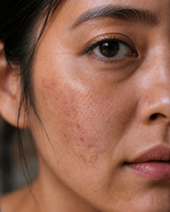
Chronically elevated cortisol may accelerate visible skin aging. New research also suggests that sensory nerves help regulate how skin ages, and both processes depend on energy-intensive cellular work.
Pontos principais
- Cortisol is necessary, but prolonged stress signaling can interfere with skin maintenance and recovery.
- Human studies link psychological stress with slower barrier recovery, greater water loss, and rougher-looking skin.
- Glucocorticoid signaling can reduce collagen production in laboratory-grown human dermal fibroblasts.
- New research suggests that sensory nerves communicate with fibroblasts and help regulate collagen maintenance.
- Sleep, UV protection, movement, nutrition, and consistent skin care remain the most practical forms of support.
Your skin can look different after a difficult week. It may feel drier, appear duller, or show fine lines that seem more noticeable.
Some of this is temporary. Poor sleep, dehydration, facial tension, and disrupted routines can quickly affect appearance. But stress also creates biological signals that reach the skin itself. Cortisol is one of the most important.
Scientists are also learning that skin aging is not controlled only by skin cells. A 2026 study published in Cell found that sensory nerves extending into the skin communicate with collagen-producing fibroblasts. When that communication was disrupted in experimental models, fibroblast senescence increased and collagen declined.
Together, these findings show that skin aging involves hormones, nerves, fibroblasts, the skin barrier, the extracellular matrix, and the cellular energy systems that keep them working.
Cortisol Is Useful Until the Signal Stays On Too Long
Cortisol is often called the stress hormone, but it is not inherently harmful. Your body needs it to regulate metabolism, immune activity, blood pressure, and the response to physical or psychological demand.
Cortisol also follows a daily rhythm. Levels normally rise around waking and gradually fall toward the evening. A temporary increase during a demanding situation helps mobilize energy.
The concern is prolonged exposure, repeated stress without adequate recovery, or disruption of that normal rhythm. Under those conditions, a signal designed for short-term adaptation may begin to compete with tissue maintenance.
Skin is sensitive to this tradeoff because it is always working. Keratinocytes maintain the outer barrier. Fibroblasts support the collagen-rich matrix. Immune cells monitor the tissue. Blood vessels deliver oxygen and nutrients. Nerves sense the environment and communicate with nearby cells.
All of this requires coordination and energy. For a wider look at how prolonged stress affects cellular energy and recovery, read Cellular Health and Stress Management.
Stress Can Slow Skin-Barrier Recovery
The outer skin barrier keeps water in while helping keep irritants and microorganisms out. It depends on organized layers of cells, lipids, structural proteins, and immune signals.
Human experiments suggest that psychological stress can temporarily slow barrier repair. In a randomized study of 85 healthy adults, a laboratory stress task delayed recovery after controlled tape stripping. The difference was measurable within hours.
One stressful event does not permanently age the skin. The study does show that a mental stressor can affect a physical skin function.
When stress is frequent and recovery is incomplete, the skin may spend more time in a less resilient state. This may contribute to dryness, sensitivity, roughness, and greater water loss. Poor sleep and disrupted skin-care habits can add to the effect.
This is why stress-related skin changes are rarely caused by cortisol alone. Hormonal signaling and everyday behavior often move together.
Cortisol Signaling Can Affect Fibroblasts and Collagen
Fibroblasts are the main support cells in the dermis. They produce and remodel collagen and other components of the extracellular matrix that help give skin strength and firmness. To learn more about this relationship, read Mitochondria, Collagen, and Skin Structure.
These cells respond to UV exposure, inflammation, oxidative stress, hormones, mechanical tension, nutrients, and cellular energy status.
Glucocorticoids can act directly on fibroblasts through the glucocorticoid receptor. In a 2023 laboratory study, researchers exposed human dermal fibroblasts to dexamethasone, a synthetic glucocorticoid. The treatment reduced type I collagen synthesis.
This was a cell-culture study using a medication, not proof that everyday stress directly creates wrinkles. Still, it identifies a plausible pathway through which prolonged glucocorticoid exposure could influence collagen maintenance.
A small 2025 human study compared women reporting mild stress with women reporting moderate chronic stress. The more stressed group had greater transepidermal water loss, lower measured antioxidant capacity, and rougher skin across several measurements.
The study included only 36 participants and grouped them by perceived stress, not by a diagnosed cortisol disorder. It cannot prove that stress caused the differences.
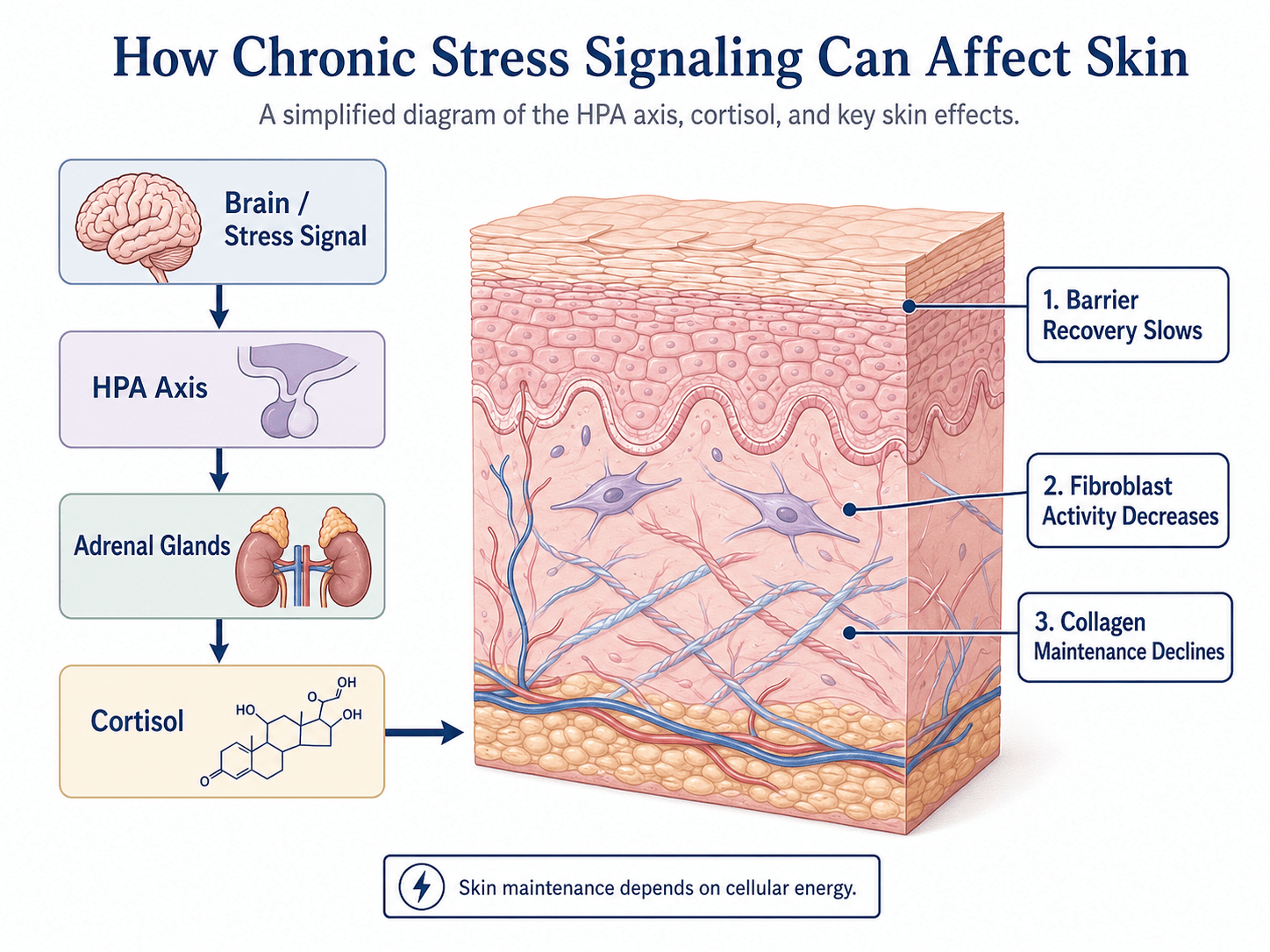
The careful conclusion is that chronic perceived stress is associated with measurable changes in skin texture and barrier function, while laboratory studies provide possible biological explanations.
Sensory Nerves maio Help Regulate Skin Aging
The nervous system has usually been discussed in skin care through sensation: pain, touch, temperature, pressure, and itch.
New research suggests that skin nerves also help maintain the tissue around them.
In the 2026 Cell study, researchers examined skin-innervating neurons that release glutamate. Their nerve fibers frequently contacted dermal fibroblasts. When the skin was experimentally denervated, collagen loss and signs of aging increased.
The researchers also studied neurofilament heavy chain, or NEFH, a neuronal structural protein that declined with age. Removing the corresponding gene from glutamatergic neurons increased fibroblast senescence and collagen loss in experimental models.
The proposed pathway involves neuronal NEFH, the enzyme CDK5, glutamate released by the nerve, and a glutamate transporter in fibroblasts. Together, this signaling system appeared to influence whether fibroblasts remained functional or moved toward senescence.
This does not mean that consuming or applying glutamate will make skin look younger. Most of the causal experiments were performed in mice and cultured cells. Human clinical research is still needed.
The important finding is broader: sensory nerves appear to participate directly in skin maintenance.
Cellular Energy Connects the Two Pathways
Hormonal stress and nerve signaling seem like separate subjects but both depend on cellular energy.
Fibroblasts need ATP to produce proteins, organize collagen, and remodel the extracellular matrix. Nerve cells also require substantial energy to maintain electrical gradients, transport materials, and release chemical signals.
Mitochondria help provide that energy and participate in stress-response signaling. When mitochondrial function is strained, skin cells may have less capacity to meet energy demand while managing oxidative stress.
Research in human dermal fibroblasts has linked UVA exposure with lower mitochondrial quality, reduced ATP, and impaired production of type I collagen and fibrillin-1. This explains why cellular energy belongs in the wider explanation.
Skin appearance reflects whether cells can communicate, maintain barriers, produce structural proteins, and recover from repeated environmental and hormonal stress.
What This Means in Real Life
Stress is one contributor to skin aging, not the whole explanation. Genetics, age, UV exposure, smoking, pollution, hormones, facial movement, nutrition, hydration, sleep, and skin-care habits all matter.
The goal is not to eliminate cortisol. It is to help the stress response return to baseline when the demand has passed.
Protect Sleep Timing
A regular wake time, enough sleep opportunity, morning light, and lower light exposure late at night help reinforce the daily rhythms that regulate cortisol and tissue recovery.
Reduce Unnecessary UV Exposure
UV exposure remains one of the clearest modifiable drivers of visible skin aging. Use shade, protective clothing, sensible timing, and sunscreen when exposure is prolonged or intense.
Support the Skin Barrier
Use a gentle cleanser and an appropriate moisturizer. Avoid repeatedly over-exfoliating skin that is already dry, sensitive, or irritated.
Move and Eat for Recovery
Physical activity supports sleep, circulation, metabolic regulation, and stress recovery. Adequate protein, essential fats, vitamins, minerals, and varied plant foods provide raw materials for normal tissue maintenance.
Do Not Diagnose High Cortisol From Appearance
Puffiness, breakouts, dryness, or tired-looking skin do not establish a cortisol disorder. True cortisol excess requires medical evaluation.
Persistent symptoms such as easy bruising, marked muscle weakness, wide purple stretch marks, or unusual changes in fat distribution should be discussed with a clinician.
Where Mitozz and Mitozz RS Fit In
The science of stress-related skin aging points to two connected needs: supporting the body’s internal energy systems and caring for the skin directly at the surface.
Mitozz is designed for the internal side of that equation. It contains 98% pure (−)-epicatechin, a compound studied in relation to mitochondrial signaling and cellular energy. Because fibroblasts, sensory nerves, and other skin cells depend on steady energy to communicate, maintain tissue, and respond to stress, mitochondrial health belongs in the broader conversation about how skin ages over time.
Mitozz RS addresses the skin more directly. It is a topical cosmetic serum formulated with (−)-epicatechin and ingredients that support hydration, conditioning, and the visible appearance of the skin. Used consistently, it is designed to help skin look smoother, more even, and well cared for.
Together, Mitozz and Mitozz RS reflect a two-part approach: supporting cellular energy from within while providing targeted cosmetic care on the outside. They do not replace sleep, stress recovery, UV protection, nutrition, or a consistent skin-care routine. They are designed to fit alongside those foundations.
Conclusion
The relationship between stress and skin aging goes beyond simply looking tired.
Prolonged stress signaling may affect barrier recovery, fibroblast activity, collagen maintenance, and the skin’s response to daily strain. New research adds sensory nerves to the picture, showing that they may communicate directly with fibroblasts and help regulate collagen homeostasis.
The most useful response is not a cortisol detox or a single anti-aging ingredient. It is to support the conditions in which skin cells can keep doing their normal work.
Healthy-looking skin reflects more than what’s on the surface. It reflects how well the whole tissue communicates, maintains itself, and meets its daily energy demands. Improving mitochondrial health helps skin cells maintain the energy, repair capacity, and resilience needed to support healthier-looking skin over time.
References
- Wang, Z., Jin, X., Wu, Y., et al. (2026). Skin-innervating glutamatergic neurons modulate aging. Cell.
- Pujos, M., Chamayou-Robert, C., Parat, M., et al. (2025). Impact of chronic moderate psychological stress on skin aging: Exploratory clinical study and cellular functioning. Journal of Cosmetic Dermatology, 24, e16634.
- Robles, T. F. (2007). Stress, social support, and delayed skin barrier recovery. Psychosomatic Medicine, 69(8), 807–815.
- Choi, D., Kang, W., Park, S., Son, B., and Park, T. (2023). Identification of glucocorticoid receptor target genes that potentially inhibit collagen synthesis in human dermal fibroblasts. Biomolecules, 13(6), 978.
- Zhao, X., Psarianos, P., Ghoraie, L. S., et al. (2019). Metabolic regulation of dermal fibroblasts contributes to skin extracellular matrix homeostasis and fibrosis. Nature Metabolism, 1, 147–157.
- Katsuyama, Y., Yamawaki, Y., Sato, Y., et al. (2022). Decreased mitochondrial function in UVA-irradiated dermal fibroblasts causes insufficient formation of type I collagen and fibrillin-1 fibers. Journal of Dermatological Science, 108(1), 22–29.