
Mitocôndrias e longevidade
The cellular foundation of healthspan.
Healthy aging depends not only on how long we live, but on how well our cells continue to produce energy, adapt, and repair themselves over time.
Living longer is only part of the goal.
Scientific and clinical attention is increasingly focused on healthspan: the portion of life spent with strong physical, cognitive, and metabolic function.
Healthspan reflects the capacity to maintain vitality, independence, and quality of life well into advanced age, with less burden from chronic disease and functional decline.
Meaningful extensions in healthspan depend on the biological processes that govern how cells produce energy, repair damage, and remain resilient as demands change over time.
Mitochondrial function connects energy production, cellular maintenance, and the ability to adapt.
Mitochondria are not the only biological regulators of aging, but they interact with many of the processes associated with healthspan, including nutrient sensing, cellular quality control, oxidative signaling, senescence, and communication between cells.
Mitochondria do more than produce energy.
Mitochondria are dynamic cellular structures that help coordinate energy conversion, metabolic adaptation, signaling, and the renewal of the mitochondrial network.
Because mitochondria serve several connected roles, changes in their function can affect how well cells and tissues respond to rising stress and energy demands over time.
What changes in mitochondria as we age?
Aging is associated with changes in mitochondrial energy production, quality control, signaling, and the capacity to respond to stress. Together, these changes may reduce cellular resilience over time.
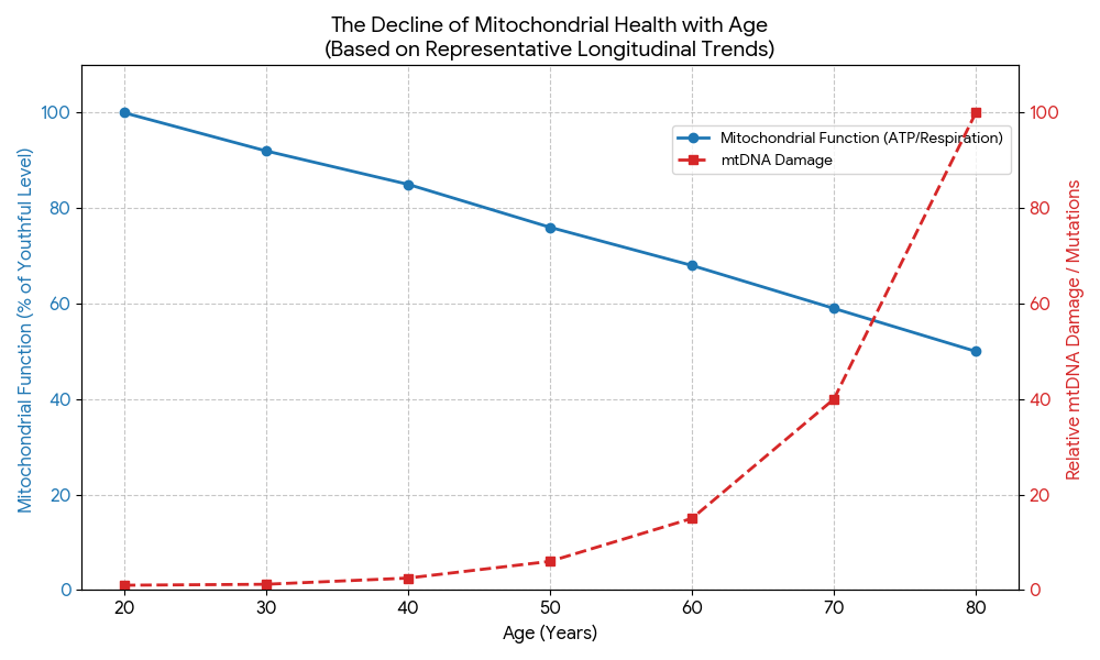
Because tissues differ in their energy needs and mitochondrial demands, age-related changes may become more noticeable in some parts of the body than in others.
Why high-energy tissues may feel the effects first.
Every tissue depends on mitochondria, but some require especially large and continuous supplies of cellular energy. The examples below represent tissues and systems in which maintaining mitochondrial capacity may be especially important for ongoing function, repair, and adaptation.
Brain
The brain requires a continuous supply of energy to maintain electrical signaling, communication between neurons, memory, and cognitive function. Limited mitochondrial reserve may make it more difficult to sustain these processes under stress.
Eyes and vision
The retina and other visual tissues have high and continuous energy needs. Mitochondrial function helps support visual signaling, cellular maintenance, and the ongoing renewal required by specialized cells involved in sight.

Heart and circulation
The heart works continuously and has exceptionally high cellular energy requirements. Mitochondria also participate in processes that support vascular function, circulation, and the delivery of oxygen and nutrients throughout the body.
Reproductive tissues
Mitochondria support energy-intensive processes involved in reproductive cell function, hormone production, and egg maturation, as well as sperm movement, fertilization, and the earliest stages of development.
Skeletal muscle
Muscles rely on mitochondria to support movement, endurance, recovery, and adaptation to physical activity. Changes in mitochondrial function may reduce the energy available when muscular demand increases.
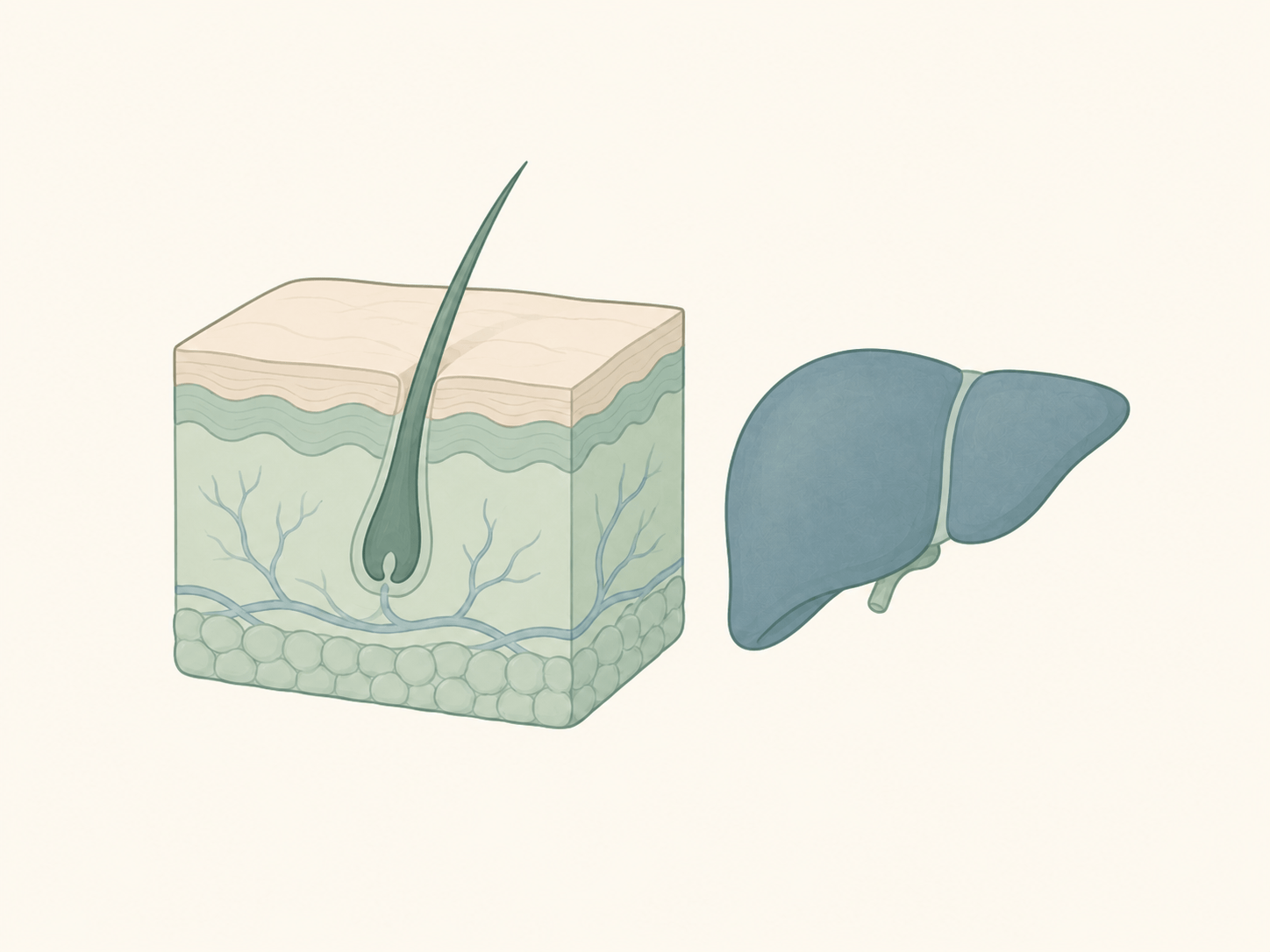
Skin and metabolic tissues
Skin depends on mitochondrial energy for renewal, repair, and responses to environmental stress. The liver and other metabolically active tissues rely on mitochondrial function for nutrient processing, fuel regulation, and metabolic adaptation. Their specific needs differ, but each relies on maintaining adequate cellular energy.
These tissues are representative examples, not an exhaustive list. Mitochondrial health supports interconnected organs and systems throughout the body and contributes to physical, cognitive, metabolic, and reproductive function across the lifespan.
Mitochondria interact with the systems that shape how cells age.
Mitochondrial function is closely connected with the systems that regulate cellular repair, stress responses, nutrient sensing, and the removal of damaged components. These connections also help explain how mitochondrial stress and inflammation can reinforce one another.
These connections help explain why mitochondrial health can influence more than cellular energy alone and why it remains central to the biology of healthspan.
Mitochondria remain responsive to how we live.
Mitochondrial function changes with age, but mitochondria continue responding to signals from movement, sleep, nutrition, recovery, and metabolic demand. No single habit controls aging, yet the signals cells receive repeatedly can influence energy capacity, maintenance, and resilience over time.
Movement
Learn how exercise supports mitochondrial adaptation, energy capacity, and recovery.
Sleep
Understand what happens to mitochondrial energy and maintenance when sleep is consistently cut short.
Nutrition
Explore how food timing, nutrient availability, and fasting influence cellular energy.
Stress and recovery
See how chronic stress can redirect cellular energy away from repair and recovery.
Two formulations built around high-purity (−)-epicatechin.
Movement, sleep, nutrition, recovery, and supplementation are all ways people support mitochondrial health. The products below provide distinct daily and topical formulations.

Mitozz
A defined, single-compound formula delivering 98% pure (−)-epicatechin to support healthy mitochondrial function, normal cellular energy, and the systems cells use to adapt to changing demands.*

Mitozz
A lightweight serum formulated with 98% pure (−)-epicatechin to hydrate and condition the skin while improving the appearance of fine lines, wrinkles, visible blemishes, and uneven-looking tone..
Scientific appendix
Scientific references
Review the research informing this educational overview.
Scientific references
Review the research informing this educational overview.
Sources are grouped by the areas of mitochondrial biology and healthspan discussed throughout the page.
Foundations of mitochondrial aging
5 sources- López-Otín, C., Blasco, M. A., Partridge, L., Serrano, M., & Kroemer, G. (2023). Hallmarks of aging: An expanding universe. Cell, 186(2), 243–278.
- Sun, N., Youle, R. J., & Finkel, T. (2016). The mitochondrial basis of aging. Molecular Cell, 61(5), 654–666.
- Bratic, A., & Larsson, N. G. (2013). The role of mitochondria in aging. Journal of Clinical Investigation, 123(3), 951–957.
- Burtscher, J., et al. (2023). Mitochondrial stress and mitokines in aging. Aging Cell, 22(2), e13770.
- Whitehall, J. C., Smith, A. L. M., & Greaves, L. C. (2023). Mitochondrial DNA mutations and ageing. Subcellular Biochemistry, 102, 77–98.
Mitochondrial quality control, signaling, and adaptation
4 sources- Youle, R. J., & Narendra, D. P. (2011). Mechanisms of mitophagy. Nature Reviews Molecular Cell Biology, 12(1), 9–14.
- Chandel, N. S. (2014). Mitochondria as signaling organelles. BMC Biology, 12, 34.
- Ristow, M., & Schmeisser, K. (2014). Mitohormesis: Promoting health and lifespan by increased levels of reactive oxygen species (ROS). Dose-Response, 12(2), 288–341.
- Wu, Z., et al. (1999). Mechanisms controlling mitochondrial biogenesis and respiration through the thermogenic coactivator PGC-1. Cell, 98(1), 115–124.
Lifestyle and mitochondrial resilience
5 sources- Seo, D. Y., et al. (2016). Age-related changes in skeletal muscle mitochondria: The role of exercise. Integrative Medicine Research, 5(3), 182–186.
- Melhuish Beaupre, L. M., Brown, G. M., Braganza, N. A., Kennedy, J. L., & Gonçalves, V. F. (2022). Mitochondria’s role in sleep: Novel insights from sleep deprivation and restriction studies. World Journal of Biological Psychiatry, 23(1), 1–13.
- Picard, M., & McEwen, B. S. (2018). Psychological stress and mitochondria: A systematic review. Psychosomatic Medicine, 80(2), 141–153.
- Fontana, L., & Partridge, L. (2015). Promoting health and longevity through diet: From model organisms to humans. Cell, 161(1), 106–118.
- de Cabo, R., & Mattson, M. P. (2019). Effects of intermittent fasting on health, aging, and disease. New England Journal of Medicine, 381(26), 2541–2551.
Mitochondria across high-energy tissues
7 sources- Mattson, M. P., & Arumugam, T. V. (2018). Hallmarks of brain aging: Adaptive and pathological modification by metabolic states. Cell Metabolism, 27(6), 1176–1199.
- Campello, L., et al. (2021). Aging of the retina: Molecular and metabolic turbulences and potential interventions. Annual Review of Vision Science, 7, 633–664.
- Ali, M. A., Gioscia-Ryan, R., Yang, D., Sutton, N. R., & Tyrrell, D. J. (2024). Cardiovascular aging: Spotlight on mitochondria. American Journal of Physiology–Heart and Circulatory Physiology, 326(2), H317–H333.
- Ramalho-Santos, J., & Amaral, S. (2013). Mitochondria and mammalian reproduction. Molecular and Cellular Endocrinology, 379(1–2), 74–84.
- maio-Panloup, P., et al. (2016). Ovarian ageing: The role of mitochondria in oocytes and follicles. Human Reproduction Update, 22(6), 725–743.
- Martic, I., Papaccio, F., Bellei, B., & Cavinato, M. (2023). Mitochondrial dynamics and metabolism across skin cells: Implications for skin homeostasis and aging. Frontiers in Physiology, 14, 1284410.
- Ganguly, S., et al. (2025). Aging and aging-related senescence in liver. Seminars in Liver Disease, 45(4), 549–566.
(−)-Epicatechin and mitochondrial support
3 sources- Daussin, F. N., Heyman, E., & Burelle, Y. (2021). Effects of (−)-epicatechin on mitochondria. Nutrition Reviews, 79(1), 25–41.
- Nogueira, L., et al. (2011). (−)-Epicatechin enhances fatigue resistance and oxidative capacity in mouse muscle. The Journal of Physiology, 589(18), 4615–4631.
- McDonald, C. M., et al. (2021). (−)-Epicatechin induces mitochondrial biogenesis and markers of muscle regeneration in adults with Becker muscular dystrophy. Muscle & Nerve, 63(2), 239–249.
This selected bibliography includes the sources most directly connected with the subjects covered on this page. It is not an exhaustive review of mitochondrial or longevity research.
* These statements have not been evaluated by the Food and Drug Administration. This product is not intended to diagnose, treat, cure, or prevent any disease.
Information presented on this page is provided for general educational purposes and is not a substitute for professional medical advice, diagnosis, or treatment.